Educational disclaimer. This article is general biological reference only. It is not medical advice, not a clinical guide, and does not describe diagnosis or treatment of human disease. If you suspect any eye or neurological condition, seek qualified medical attention promptly.
There is almost certainly Acanthamoeba in your tap water. It is in the soil in your garden, in ocean sediment, in dust particles in the air, in rivers, in hot tubs, in swimming pools, in the biofilm that lines plumbing. It has been collected from Antarctica, from deep-sea sediment, and from swabs taken inside hospital ventilation systems. Unlike the headline-grabbing Naegleria fowleri, Acanthamoeba is not a rare organism in specialized habitats. It is one of the most globally distributed, environmentally resilient free-living amoebae in the microbial world.
And it is also, under specific circumstances, capable of infecting the human eye and brain.
What kind of organism is Acanthamoeba?
Acanthamoeba belongs to the class Discosea, order Centramoebida. It is classified among the Amoebozoa — a diverse group that includes everything from garden-variety soil amoebae to the social slime molds. There are at least 24 recognized species of Acanthamoeba, traditionally grouped into genotypes (T1 through T22) based on the variable region of the 18S ribosomal RNA gene. The most clinically relevant genotypes are T4 (by far the most common worldwide), T3, T6, T11, and several others.
Despite its clinical significance, Acanthamoeba is not a parasite in the conventional sense. It does not need any host organism to complete its life cycle. It feeds entirely on bacteria, algae, yeast, and organic particles in its environment — and can go months or years without any of them, thanks to a remarkably resilient resting form.
Two life stages, one armored form
Unlike Naegleria fowleri, which has three morphological stages (trophozoite, cyst, flagellate), Acanthamoeba has only two:
Trophozoite
The active, feeding form. Acanthamoeba trophozoites are 25 to 40 micrometers across and are recognized by their characteristic acanthopodia — long, thin, spine-like projections extending from the cell surface that give the genus its name (akantha = thorn, in Greek). These are not feeding structures but are involved in locomotion and surface adhesion. The trophozoite moves slowly using broad pseudopodia and engulfs food by phagocytosis.
Cyst
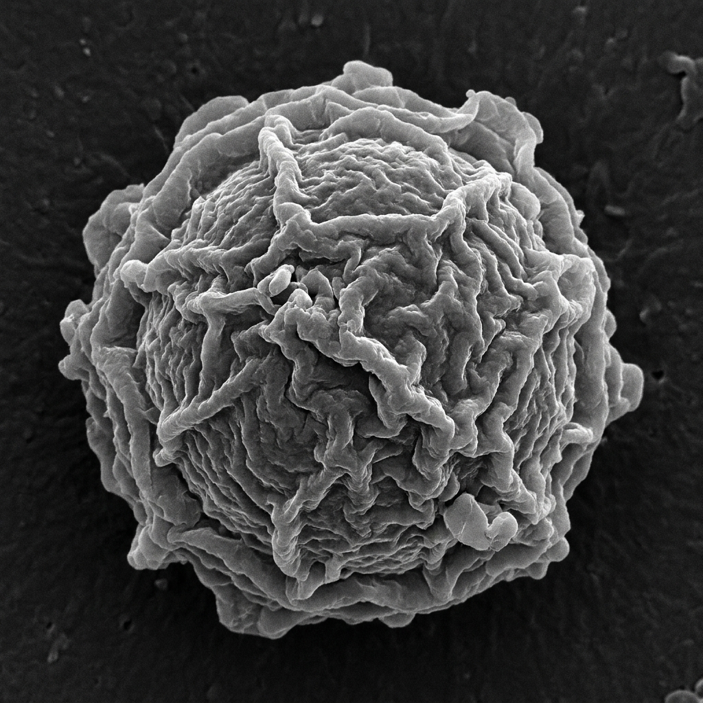
When conditions become adverse — cold temperature, dryness, nutrient depletion, high osmolarity — the trophozoite encysts. The resulting cyst is structurally remarkable: it has a double wall, consisting of an outer ectocyst (typically stellate or polygonal, species-dependent) and an inner endocyst (usually spherical). Between these walls there is a gap crossed by connecting extensions that differ between species and are used in identification.
The cyst’s double wall is composed largely of cellulose and proteins, rendering it extraordinarily resistant to:
- Disinfectants, including chlorine at concentrations used in most water-treatment systems.
- Desiccation — cysts can survive completely dry for months to years.
- Temperature extremes — from near-freezing to well above 50 °C in some conditions.
- UV radiation — at doses that sterilize most bacteria.
- Many antiparasitic compounds.
This resistance is why Acanthamoeba is found in environments that are nominally treated or disinfected. A standard swimming pool is chlorinated enough to kill most pathogens, but Acanthamoeba cysts persist, waiting to excyst if conditions warm.
The Trojan horse: Acanthamoeba as a bacterial carrier
One of the most biologically interesting aspects of Acanthamoeba ecology is its relationship with certain bacteria. Many bacteria are simply digested when the amoeba engulfs them. But some have evolved to resist digestion inside the phagosome — and in doing so, they gain a protective niche inside a cell that is itself extremely resistant to environmental destruction.
Well-documented intracellular passengers include:
- Legionella pneumophila — the bacterium responsible for Legionnaires’ disease. Legionella is unable to infect humans directly through the water supply at normal concentrations; instead, it replicates inside Acanthamoeba in water systems (cooling towers, hot water tanks, hospital plumbing), amplifying massively before being aerosolized into air that humans breathe.
- Mycobacterium spp. — including some non-tuberculous mycobacteria that can persist inside amoebae in tap water and plumbing biofilms.
- Various emerging opportunistic pathogens that have been found to survive amoeba phagocytosis and replicate intracellularly.
This makes Acanthamoeba a “Trojan horse” in water microbiology — a vehicle that can concentrate and protect dangerous bacteria inside a cyst that survives treatment, then release viable bacteria when environmental conditions change. It is one of the reasons modern drinking-water systems are designed with specific protocols targeting amoeba-associated bacterial risks, not just direct bacterial contamination.
How Acanthamoeba reaches human tissue
Acanthamoeba is not adapted to infect humans. It has no specialized invasion mechanisms for breaking through intact skin or mucosa in healthy individuals. The two main disease presentations — Acanthamoeba keratitis (AK) and granulomatous amoebic encephalitis (GAE) — arise through very different routes and in very different risk populations.
Route 1: The eye (Acanthamoeba keratitis)
The cornea of the eye is one of the few places where Acanthamoeba trophozoites can adhere to and penetrate tissue in an otherwise healthy, immunocompetent person. The biological reason involves specific binding between Acanthamoeba mannose-binding proteins and mannose-conjugated glycoproteins expressed on corneal epithelial cells.
Risk is strongly associated with contact lens wear, particularly when lenses are:
- Rinsed in or stored in tap water rather than sterile contact lens solution.
- Worn while swimming, showering, or in hot tubs.
- Kept in contaminated lens cases (biofilm inside cases is a documented reservoir).
- Worn for extended periods without appropriate disinfection protocols.
The eye of a contact lens wearer offers Acanthamoeba trophozoites optimal conditions: a warm, moist, low-competition surface, somewhat disrupted by the mechanical abrasion of lens wear, with a specific molecular handle (mannose receptors) the amoeba recognizes. AK does not require immunosuppression.
Route 2: The brain (Granulomatous Amoebic Encephalitis)
GAE is a very different clinical entity and follows a different biological route. It occurs almost exclusively in immunocompromised individuals: people with advanced HIV/AIDS, organ-transplant recipients on immunosuppressive therapy, and individuals on high-dose corticosteroid treatment.
The proposed route is hematogenous — the amoeba (or its antigens) reaches the bloodstream from a skin wound, the lungs, or another primary site of colonization, and crosses into the brain via the blood-brain barrier. This is fundamentally different from Naegleria fowleri, which travels the olfactory nerve from the nasal cavity. Acanthamoeba GAE is a slow, subacute process, not the rapid fulminant course of Naegleria PAM.
A third rarer condition, disseminated cutaneous acanthamoebiasis, produces skin nodules and ulcers in immunocompromised patients; it sometimes precedes neurological involvement.
Environmental ubiquity and what it means
Because Acanthamoeba is so universally present, most humans are exposed to it constantly — in the water we use, the air we breathe, the soil we touch. Most of the time, nothing happens. The immune system of a healthy person clears the occasional trophozoite that crosses a mucous membrane without difficulty. Most immunocompetent people carry Acanthamoeba-specific antibodies detectable in serum and tears — silent evidence of exposure and immune clearance that never produced symptoms.
This immunological background is itself informative: it tells us that low-level exposure is essentially universal, which means the organism’s ecological success is not matched by epidemiological danger under normal circumstances.
Practical prevention
Because the two disease presentations have different risk factors, prevention focuses on distinct behaviors:
For Acanthamoeba keratitis (relevant to contact lens wearers):
- Never rinse, store, or clean contact lenses with tap water. Use only sterile, commercially prepared contact lens solution.
- Do not wear contact lenses while swimming, in hot tubs, or in the shower.
- Replace lens cases regularly and disinfect them according to manufacturer guidelines.
- Remove contact lenses before sleeping unless prescribed extended-wear lenses by a qualified practitioner.
For GAE (relevant during periods of immunosuppression, in consultation with medical professionals):
- Follow guidance from your treating physician regarding water exposure and wound care.
- General wound hygiene — clean, covered wounds — reduces skin as an entry point.
These are general behavioral precautions derived from what is understood about Acanthamoeba biology. They are not medical advice, and anyone with specific health concerns should consult a qualified healthcare provider.
Why it matters
Acanthamoeba sits at an unusual intersection of ecology, public-health microbiology, and opportunistic infection. Understanding it requires thinking simultaneously about:
- Environmental microbiology — how an extraordinarily resilient cyst persists through disinfection and travels globally in water systems.
- Microbial ecology — how Acanthamoeba’s role as a bacterial predator paradoxically amplifies the survival and virulence of certain pathogenic bacteria inside water infrastructure.
- Host-parasite evolution — how an organism with no evolutionary investment in infecting vertebrates can, by chance of molecular surface chemistry, colonize a corneal epithelium or, in the absence of immune defense, a brain.
The lesson is not “be afraid of tap water.” It is something more biologically nuanced: the ecology of an organism and its medical relevance are not always proportional. Acanthamoeba is globally ubiquitous and almost never causes disease. The small fraction of exposures that do produce pathology arise from a collision of opportunistic binding ability, a specific anatomical vulnerability (the contact-lens-worn cornea), or the removal of the immune defense that usually keeps free-living amoebae exactly where they belong — outside the body.
In a world where microbial diversity is almost incomprehensibly vast and most of it never intersects with human disease, Acanthamoeba is a modest, well-documented reminder of how thin and context-dependent the boundary between environmental microorganism and clinical pathogen really is.
Sources and further reading
- Centers for Disease Control and Prevention. Acanthamoeba Infection. cdc.gov/parasites/acanthamoeba
- Visvesvara GS, Moura H, Schuster FL. “Pathogenic and opportunistic free-living amoebae.” FEMS Immunology and Medical Microbiology 50(1):1–26. 2007.
- Garajová M et al. “Acanthamoeba keratitis: risk factors and prevention.” Expert Review of Anti-infective Therapy 18(9):905–914. 2020.
- Thomas PA, Geraldine P. “Infectious keratitis.” Current Opinion in Infectious Diseases 20(2):129–141. 2007.