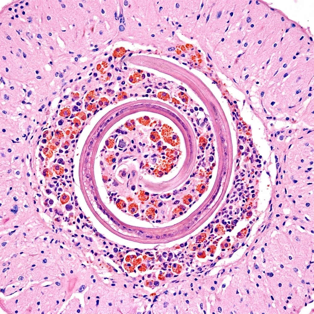
Educational disclaimer. This article is general biological reference only. It is not medical advice, not a clinical guide, and does not describe diagnosis or treatment of human disease. If you suspect any parasitic or neurological condition, seek qualified medical attention promptly.
Angiostrongylus cantonensis, commonly called the rat lungworm, is a parasitic nematode whose adult form lives in the pulmonary arteries of rats. In its normal life cycle it never touches a human. When it does — usually through a slice of contaminated lettuce or a raw snail eaten on a dare — the larva ends up in the wrong species and travels to the wrong organ. It is now the leading global cause of eosinophilic meningitis, with confirmed cases on every inhabited continent.
The worm was first described in 1935 by the Chinese parasitologist H.T. Chen, who recovered it from rats in Canton (modern Guangzhou) — hence cantonensis. The first confirmed human case appeared in Taiwan in 1944. In the decades since, A. cantonensis has spread from Southeast Asia and the Pacific to Hawaii, the Caribbean, Australia, southern Europe, and the southern United States, hitchhiking inside its rodent and mollusk hosts as global trade has expanded.
A two-host life cycle (with detours)
The worm’s natural life cycle alternates between a vertebrate definitive host and an invertebrate intermediate host:
- Definitive host — rats. Adult worms (males ~20 mm, females ~25–35 mm) live coiled in the pulmonary arteries of rats, primarily Rattus rattus and Rattus norvegicus. Females release eggs that hatch into first-stage larvae (L1) inside the rat’s lungs. The larvae are coughed up, swallowed, and passed in the rat’s feces.
- Intermediate host — snails and slugs. L1 larvae penetrate or are ingested by terrestrial and freshwater gastropods. Inside the mollusk, they develop through two molts into the infective third-stage larva (L3) over 12–14 days. The giant African land snail (Lissachatina fulica, formerly Achatina fulica) is famously efficient at carrying L3 larvae and has driven multiple outbreaks where it has invaded.
- Back to rats. A rat eats an infected snail or slug. The L3 larva penetrates the gut wall, enters the bloodstream, and migrates to the brain, where it spends about two weeks maturing through L4 and young-adult stages. From the brain it returns via the venous system to the pulmonary arteries, matures fully, mates, and the cycle restarts.
Several other animals can carry L3 larvae without further development — what parasitologists call paratenic hosts. These include freshwater prawns and crabs, frogs, planarian flatworms, monitor lizards, and centipedes. Eating a paratenic host raw, or even contaminated produce that has had slug or snail slime on it, can transmit the parasite onward.
How humans get infected
Humans become accidental hosts in the same way rats normally do — by ingesting L3 larvae — but the most common routes look very different from a rodent’s diet:
- Eating raw or undercooked snails or slugs (a documented route in cultures where these are food or, more commonly, in children eating them as a dare).
- Eating raw freshwater prawns, crabs, or frog flesh in regions where these are local dishes.
- Eating contaminated leafy vegetables — lettuce, cabbage, watercress — where a tiny slug or snail has crawled across the leaves and left infectious larvae behind in the mucus trail.
- Drinking water contaminated with snail mucus or carrying free L3 larvae.
Famous outbreaks include a 2000 cluster in Jamaica linked to Caesar salad, multiple ongoing cases in Hawaii tied to contaminated produce and rosemary salad mixes, and Mainland US cases reported from Florida, Texas, and Louisiana as climate change extends the range of suitable host snails northward.
What the larva does inside a human
Once swallowed, the L3 larva penetrates the gut, enters the bloodstream, and follows roughly the same migration path it would in a rat — straight to the central nervous system. In humans, however, the worm cannot complete its journey back to the lungs. It is stranded in the brain, meninges, and occasionally the spinal cord or eye, where it grows for a few weeks before dying.
The result is eosinophilic meningitis: an intense inflammatory reaction characterized by:
- Severe headache, often the most prominent symptom and frequently described as the worst the patient has experienced.
- Neck stiffness and photophobia, mimicking bacterial or viral meningitis.
- Paresthesias — abnormal skin sensations — sometimes in unusual distributions as the larva passes through specific nerve roots.
- Nausea, vomiting, low-grade fever.
- In severe cases: cranial nerve palsies, ataxia, coma, and death.
The defining laboratory feature is a lumbar puncture showing more than 10% eosinophils in the cerebrospinal fluid (often much higher), combined with elevated CSF protein and a normal or slightly low glucose. Peripheral blood eosinophilia is also common but is less specific.
Diagnosis: a clinical puzzle
A. cantonensis is rarely recovered from a living patient — the larva is small, deep in the CNS, and almost never appears in CSF samples in routine clinical settings. Diagnosis is therefore based on:
- Clinical pattern: eosinophilic meningitis with a compatible exposure history.
- CSF eosinophilia above 10% of total CSF leukocytes.
- Serology and PCR: real-time PCR for A. cantonensis DNA in CSF, now offered by the CDC and a small number of reference laboratories, is the most reliable confirmatory test.
- MRI findings: nonspecific but may show punctate enhancing lesions, leptomeningeal enhancement, or worm tracks in severe cases.
Ruling out other causes of eosinophilic meningitis is essential — the differential includes Gnathostoma spinigerum, Baylisascaris procyonis, neurocysticercosis, and certain fungal infections.
Treatment is mostly anti-inflammatory
Counterintuitively, the mainstay of treatment is not anthelmintic drugs. Killing the larva inside the brain releases its contents and dramatically amplifies the inflammatory response, often worsening neurological outcomes. The current standard of care, refined through large case series in Thailand, Hawaii, and southern China, emphasizes:
- Corticosteroids (prednisolone or dexamethasone) for two to four weeks to control CNS inflammation.
- Repeated lumbar punctures to relieve elevated intracranial pressure, which by itself improves headache and outcome.
- Anthelmintic agents (albendazole) used cautiously and only under steroid cover, in selected cases — the evidence remains debated, with some recent trials showing benefit when given early and others showing no clear advantage.
Most immunocompetent patients recover fully, but neurological sequelae and rare deaths still occur, particularly in pediatric cases and in those with heavy worm burdens.
A parasite riding climate and trade
A. cantonensis is now considered an emerging pathogen. Its expansion is driven by three forces:
- Invasive intermediate hosts. The giant African land snail and several semi-slug species have invaded ports across the tropics and subtropics, dragging the worm with them.
- Warming climate. Snails survive winters they previously could not, allowing the worm to establish in regions like the southern United States, southern Europe, and northern Australia.
- Global produce trade. Contaminated leafy greens move between continents and have been implicated in cluster outbreaks in tourists and travelers far outside the worm’s historical range.
The CDC and the WHO both now treat A. cantonensis as a notifiable disease in many jurisdictions, and surveillance has improved markedly since the 2010s.
Why it matters
The rat lungworm is a case study in how a tightly co-evolved parasite-host system can produce catastrophic spillover when humans intersect with it. In rats, the worm runs an efficient brain-to-lung migration that produces no significant disease. In people, the same migration ends in eosinophilic meningitis because our anatomy and immune system are not part of the parasite’s playbook.
The story also illustrates why eosinophilic meningitis has become an important emerging-infections diagnosis. A patient with severe headache and CSF eosinophilia after a salad in Hawaii, a snail-tasting dare in Thailand, or a frog dish in Laos may be carrying a worm that left a rat’s lungs months earlier and traveled across the world inside an invasive mollusk. Angiostrongylus cantonensis is, in short, one of the clearest examples we have of the price of a globalized food chain meeting a parasite that does not respect borders.
Sources and further reading
- Centers for Disease Control and Prevention. Angiostrongylus cantonensis. cdc.gov/parasites/angiostrongylus
- Wang QP et al. “Human angiostrongyliasis.” The Lancet Infectious Diseases 8(10):621–630. 2008.
- Hochberg NS, Blackburn BG. “Eosinophilic Meningitis.” New England Journal of Medicine 389(14):1299–1313. 2023.
- Hawaii Department of Health. Rat Lungworm Disease. health.hawaii.gov/docd/disease_listing/rat-lungworm-disease