Educational disclaimer. This article is general biological reference only. It is not medical advice, not a clinical guide, and does not describe diagnosis or treatment of human disease. If you suspect any parasitic infection or neurological condition, seek qualified medical attention promptly.
In 2014, a research team at the University of Cambridge published the genome of a parasitic tapeworm larva that had been removed from the brain of a 50-year-old man after spending at least four years wandering inside his central nervous system. MRI scans across that period showed the lesion moving five centimeters from one side of his brain toward the other. The patient was the first confirmed UK case of sparganosis, caused by the larva (sparganum) of Spirometra erinaceieuropaei — a pseudophyllidean tapeworm with one of the strangest, longest, and most invasive larval stages of any parasite that infects humans.
A tapeworm with no clear loyalty to a single host
S. erinaceieuropaei belongs to the order Diphyllobothriidea, a group of tapeworms whose adults typically live in the intestines of fish-eating carnivores. The adult worm is intestinal in domestic dogs, cats, foxes, and wild felids and canids. It is not a long worm by tapeworm standards — usually under a meter — but it can produce thousands of eggs daily, which leave the host in feces.
The species name erinaceieuropaei references the European hedgehog (Erinaceus europaeus), which can also carry the parasite. Distribution is, despite the name, primarily Asian: China, South Korea, Japan, Vietnam, Thailand, and Indonesia report most human cases. Sporadic infections are documented across Europe, the Americas, Africa, and Australia.
The life cycle requires water and at least two intermediate hosts before the worm can mature:
- Eggs are shed in the feces of a definitive host and hatch in freshwater into ciliated swimming larvae called coracidia.
- First intermediate host — copepods. Tiny freshwater crustaceans (Cyclops spp.) eat the coracidia. Inside the copepod, the larva develops into a procercoid.
- Second intermediate host — almost any vertebrate that eats copepods. Frogs, snakes, fish, reptiles, birds, and mammals all qualify. The procercoid penetrates the gut and develops into a plerocercoid, also called a sparganum — a small, white, ribbon-like larva, typically a few centimeters long, that encysts in muscle, subcutaneous tissue, or visceral organs.
- Back to a definitive host. A dog, cat, or wild carnivore eats a frog, snake, or fish carrying spargana. The larva develops into an adult tapeworm in the intestine and the cycle restarts.
Humans can step in at two points: by drinking water containing infected copepods, or — much more commonly — by eating raw or undercooked second-intermediate hosts (frog meat, snake meat, certain fish dishes). In several Asian traditional-medicine practices, raw frog or snake flesh is applied as a poultice to wounds, the eye, or inflamed skin. The sparganum can crawl out of the poultice and burrow directly into the patient’s tissue — a transmission route nearly unique to Spirometra.
A migrating larva that refuses to die
Once inside a human, the sparganum behaves much as it does in any other accidental host: it migrates. Unlike adult tapeworms, it does not anchor and grow in the gut. It is a wandering juvenile that never matures. With no metabolic clock telling it to stop, and no innate compass pointing toward a definitive host’s intestine, it simply keeps moving through whatever tissue is in front of it. Documented locations include:
- Subcutaneous tissue — the most common presentation, producing slow-growing, painful, sometimes migratory lumps.
- Skeletal muscle and deep fascia.
- Visceral organs — including the abdominal cavity, pleural cavity, and pericardium.
- Eye — producing orbital sparganosis, often with severe inflammation and risk of permanent vision loss.
- Spinal cord and brain — the most dangerous form, cerebral sparganosis, with focal neurological deficits, seizures, and slowly progressive symptoms over years.
Spargana have been recovered from patients decades after the initial exposure. The 2014 UK case mapped the worm’s movement in real time using serial MRI: across four years, the larva traveled approximately 5 cm through the right hemisphere, leaving a trail of gliotic lesions visible on imaging. Histopathology after surgical removal confirmed an intact, viable larva.
How does a millimeters-to-centimeters-long flatworm survive that long inside a human brain? Genome sequencing in the same 2014 study revealed several clues. S. erinaceieuropaei carries an expanded set of detoxification genes (including unusual cytochrome P450 and glutathione transferase repertoires), giving it broad resistance to host-derived oxidative stress and to many candidate antiparasitic compounds. It also expresses immunomodulatory proteins that suppress local T-cell responses, allowing it to live alongside an active immune system without being walled off.
Symptoms and diagnosis
Sparganosis is famously variable. Symptoms depend almost entirely on where the larva happens to be:
- Cutaneous / subcutaneous sparganosis: discrete, firm, slowly migrating nodules, sometimes with localized pain or itching. Often mistaken for lipomas or other benign masses for years.
- Ocular sparganosis: pain, swelling, decreased vision, sometimes a visible worm in the conjunctiva.
- Visceral sparganosis: vague abdominal pain, mass lesions seen on imaging, occasionally pleural effusion.
- Cerebral sparganosis: headaches, seizures, focal weakness, sensory changes, and slowly progressive lesions on MRI — frequently misdiagnosed at first as primary brain tumor or stroke.
Diagnosis usually requires a combination of:
- Imaging — CT or MRI showing characteristic tunnel-like or migrating lesions.
- Serology — ELISA against Spirometra antigens, available in reference laboratories.
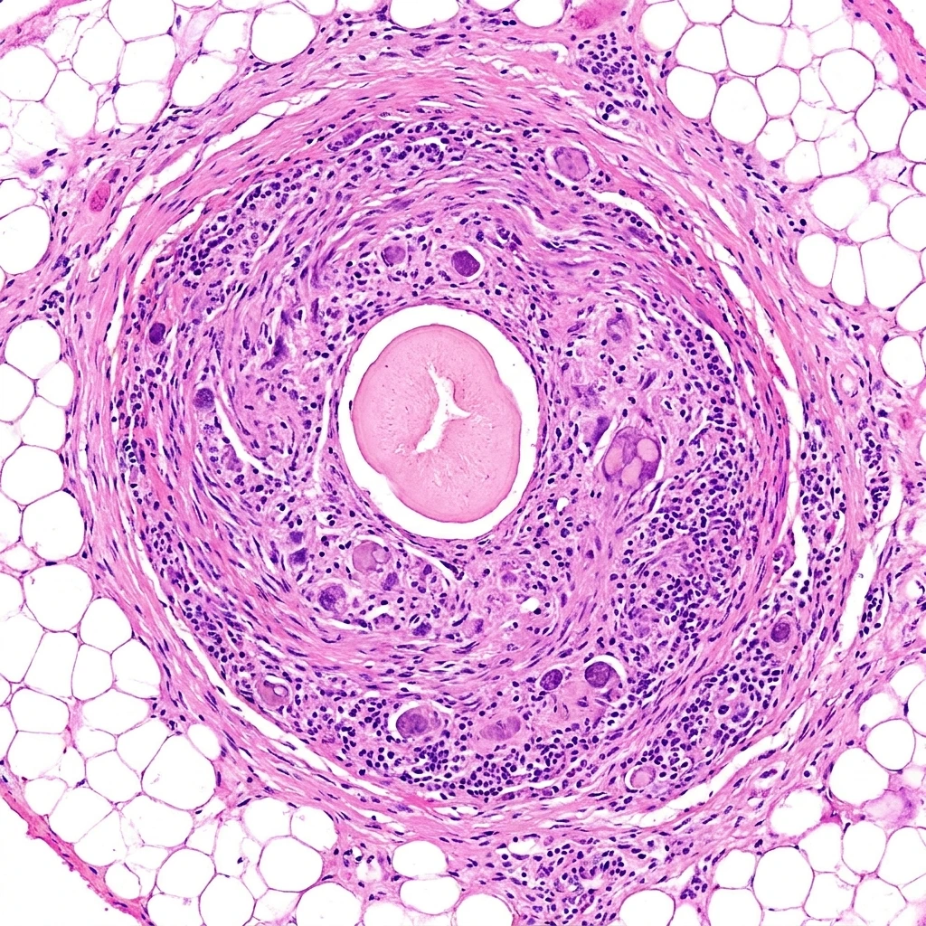
- Histopathology of excised tissue showing the characteristic plerocercoid morphology.
- Molecular confirmation — PCR of the parasite’s mitochondrial cox1 gene from biopsy material can distinguish S. erinaceieuropaei from related species such as S. proliferum, which is rarer but vastly more dangerous because it can asexually multiply inside a human host.
Treatment is mainly surgical
Pharmacological options for sparganosis are limited. Praziquantel, the standard drug for most cestode infections, has only partial efficacy against Spirometra larvae and high doses are often poorly tolerated. Albendazole has been used as an adjunct with mixed results. Killing the larva in situ in the brain or eye also risks triggering a severe inflammatory response as the dying worm releases its antigens.
For these reasons, complete surgical excision remains the first-line treatment whenever the larva can be located. In cerebral cases, neurosurgical removal under image guidance is curative if the entire worm is extracted. Where the larva cannot be reached or fully removed, prolonged drug therapy combined with corticosteroids is the next-best option, and lifelong follow-up imaging is recommended.
Why it matters
Spirometra erinaceieuropaei is one of the most extreme examples of how a parasitic larva can persist inside the wrong host. It is not particularly common — perhaps a few thousand confirmed cases worldwide in the published literature — but each case is a reminder of a few uncomfortable biological truths:
- Larvae can outlast their hosts’ patience. A worm that lives for ten or more years inside a human brain is an organism that has solved problems of immune evasion, metabolic flexibility, and tissue migration that most parasitologists assumed required a far more complex animal.
- Traditional and modern food practices intersect. Frog poultices in rural Asia and raw-snake dishes in modern restaurants both transmit the same worm. The parasite does not care about culture or century.
- Sparganum proliferum is the nightmare cousin. A close relative of S. erinaceieuropaei, the Sparganum proliferum form is capable of asexual budding inside human tissue, producing many daughter larvae from a single parent. Only a handful of confirmed cases exist, but the few documented patients have died of disseminated disease. Genomic comparison with the more common S. erinaceieuropaei may eventually reveal which genes turn a single wandering larva into a self-replicating one.
For ordinary readers, the practical lesson is simple: cook freshwater frog, snake, and fish meat thoroughly, drink only safely treated water in endemic regions, and never apply raw animal tissue to a wound. For biologists, S. erinaceieuropaei is a master class in the limits of what a “simple” flatworm can do — and a reminder that, under the right conditions, the line between infection and slow, multi-year exploration of a brain can be just one undercooked meal.
Sources and further reading
- Bennett HM et al. “Novel mitochondrial and nuclear genome sequences illuminate the phylogeny and evolutionary history of the Spirometra tapeworm.” PLOS Neglected Tropical Diseases 8(11):e3071. 2014.
- Galán-Puchades MT, Fuentes MV. “Cerebral sparganosis: from the origin of the concept to the current challenge of diagnosis.” Pathogens and Global Health 107(8):383–387. 2013.
- Li MW et al. “Sparganosis in China: an endemic zoonotic parasite.” International Journal of Infectious Diseases 14(8):e629–e635. 2011.
- Centers for Disease Control and Prevention. Sparganosis. cdc.gov/parasites/sparganosis